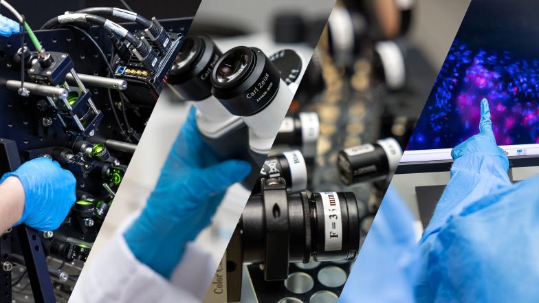
The Confocal and Light Microscope Imaging Facility is a core advanced light microscope facility on the Faculty of Health Sciences UCT campus. It offers UCT and other local researchers access to super and near-super resolution, real-time live cell and tissue imaging including a multitude of quantitative imaging and analysis techniques.
The facility is located in the Anatomy Building, level 3, rooms 3.14 & 3.18, and in Wernher & Beit South (BSLIII laboratory of the IDM), and Falmouth L3, (BSLII laboratory).
Imaging capabilities in the BSLIII:
- ImageXpressMicro Confocal, only accessible for those who complete a basic BSLIII induction course. Allows live-cell time-lapse microscopy, with sample flexibility (round, flat, tissue, etc).
- Confocal allows examination through sections. Cryosections will be available as a core facility; allows extended time lapse for samples under review.
- Light phase Transmission Microscopy with 6 fluorescent colours used. Allows live cell imaging e.g. 5-day cells imaged every 30 minutes.
- 4D viewing and 3D analyses possible, with a number of software packages available. Include 2D/3D analyses and a range of outputs within these. Allows quantification of the number of bacteria per cell and nuclei per cell. Will revolutionise how we interpret drug analyses effects.
- Zeiss Elyra (P)S.1.
- Super-resolution structured illumination microscopy (SR-SIM). To be upgraded to include Photoactivated Localisation Microscopy (PALM). Single molecule imaging.
Imaging capabilities in the BSLII:
- DeltaVision Elite high-resolution microscope, loaned from Northwestern University (NWU), USA. Will be a core facility, situated in Falmouth L3, BSLII laboratory.
Available too: Wide-field, fast high-resolution imaging that tracks dynamic movement; plus photo activation , with filters. Allows visualising of viruses using photo-activation.
Contact Suraj Parihar (BSLIII) or Caron Jacobs for bookings and further information.
